
Boling Vision Center's Cornea & Ocular Surface Center provides comprehensive corneal evaluation, medical management, and surgical care, with cornea specialists experienced in the full spectrum of conditions affecting the eye's most essential surface.
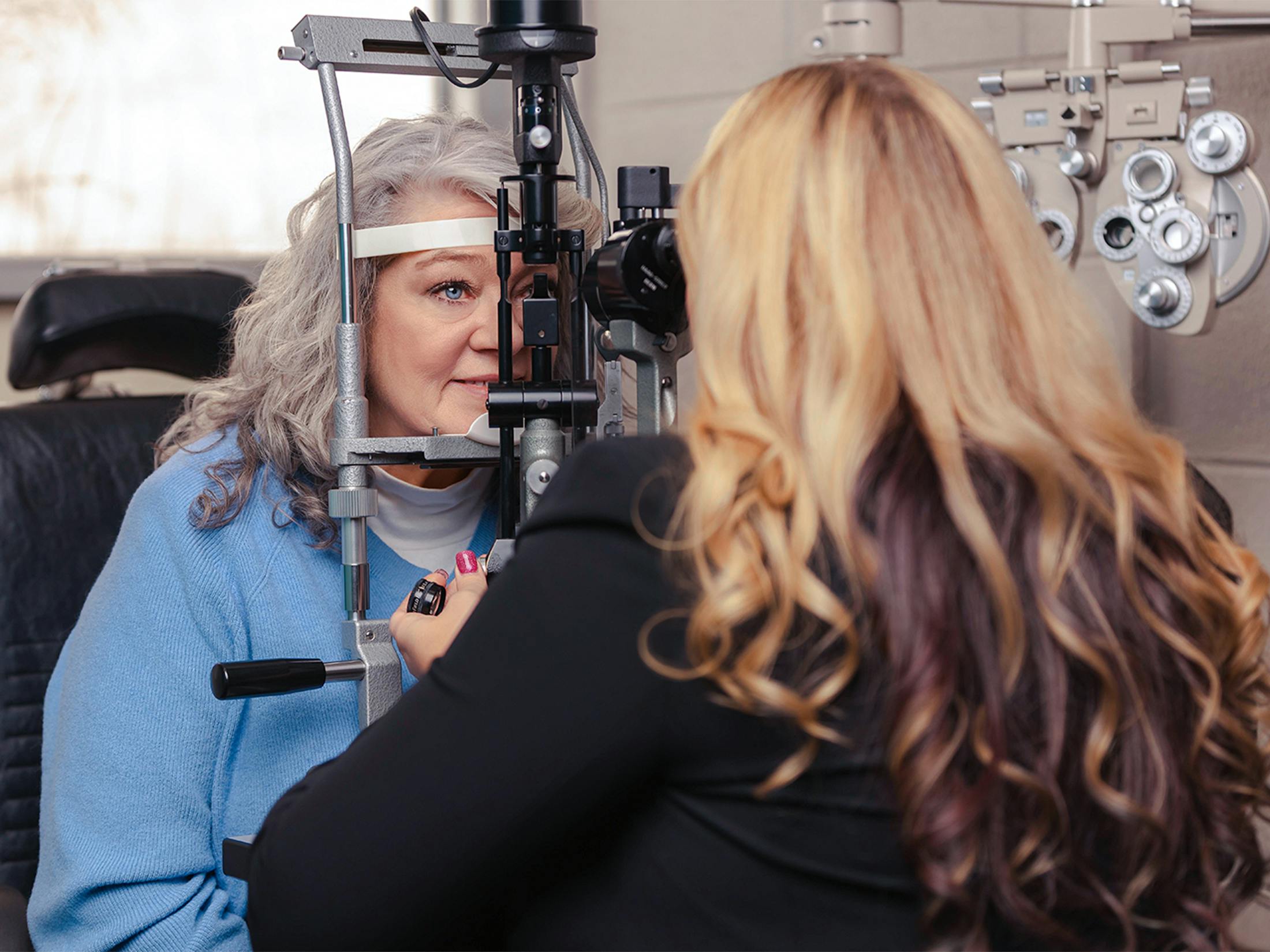



How We Diagnose Corneal Conditions
Accurate diagnosis is the foundation of effective corneal care. At Boling Vision Center, our cornea specialists use a comprehensive suite of advanced diagnostic tools.
- Corneal Topography and Tomography: Maps the curvature and thickness of the cornea in fine detail, identifying irregularities, ectatic changes, and structural abnormalities essential for diagnosing keratoconus and planning surgical treatment.
- Anterior Segment OCT: Provides cross-sectional imaging of the corneal layers, essential for characterizing scarring, dystrophies, and post-surgical changes.
- Specular Microscopy: Evaluates the health and density of the corneal endothelial cells, critical for assessing suitability for certain surgical procedures and monitoring conditions like Fuchs' dystrophy.
- Slit-Lamp Examination: A thorough slit-lamp evaluation of the cornea, conjunctiva, and eyelid margins provides direct visualization of surface health and identifies contributing conditions.