
At Boling Vision Center, our Cornea & Ocular Surface Center brings together advanced diagnostics, the full spectrum of treatment options, and providers with the clinical depth to manage everything from chronic dry eye to complex corneal disease, all under one roof in Northern Indiana.
Cornea & Ocular Surface Center
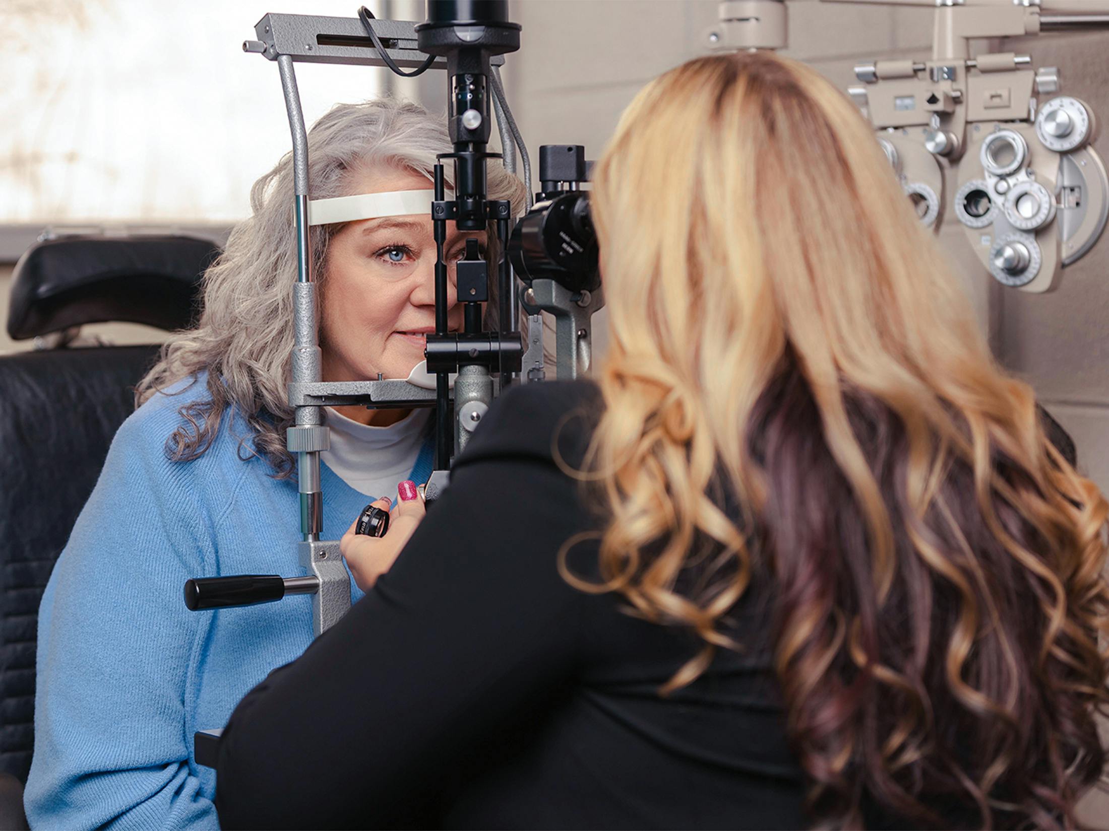

Caring for the Eye's Most Essential Surface
The cornea and ocular surface are where vision begins. Before light ever reaches the retina, it must pass through a precisely structured, continuously maintained surface system that keeps the eye clear, comfortable, and protected. When that system breaks down, whether from disease, injury, inflammation, or the effects of prior surgery, the impact on vision and quality of life can be profound.
At Boling Vision Center's Cornea & Ocular Surface Center, we specialize in evaluating and treating the full range of conditions that affect this critical part of the eye, with the diagnostic precision and therapeutic depth that complex corneal and ocular surface care demands. Schedule a consultation at any of our four Northern Indiana locations and let's take a thorough look at your corneal and ocular surface health.
Conditions We Treat
Our Cornea & Ocular Surface Center provides comprehensive evaluation and management across the full spectrum of corneal and ocular surface conditions.

Dry Eye Disease
Dry Eye Disease
One of the most common eye conditions in the world, and one of the most undertreated. Dry eye disease is rooted in a breakdown of the ocular surface system that requires accurate diagnosis and targeted treatment to address effectively, going well beyond artificial tears and one-size-fits-all drop regimens.
Meibomian Gland Dysfunction (MGD)
Meibomian Gland Dysfunction (MGD)
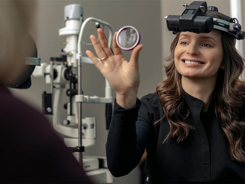
Meibomian gland dysfunction is the leading cause of evaporative dry eye, driven by blockage or dysfunction of the oil-producing glands in the eyelids that maintain the lipid layer of the tear film. MGD is extremely common, frequently underdiagnosed, and highly responsive to targeted treatment when identified and addressed appropriately.

Ocular Surface Inflammation
Ocular Surface Inflammation
Chronic inflammation and dry eye exist in a self-perpetuating cycle that keeps many guests from achieving meaningful improvement with standard treatments. Identifying and interrupting that cycle is a central focus of our ocular surface care, and addressing it directly is often what makes everything else work.
Keratoconus
Keratoconus
Keratoconus and corneal ectasia are progressive conditions in which the cornea gradually weakens and bulges into an irregular shape, producing distorted vision that becomes increasingly difficult to correct with conventional lenses. Early diagnosis and timely intervention, including corneal cross-linking, make an enormous difference in long-term outcomes.
Corneal Scarring or Trauma
Corneal Scarring or Trauma
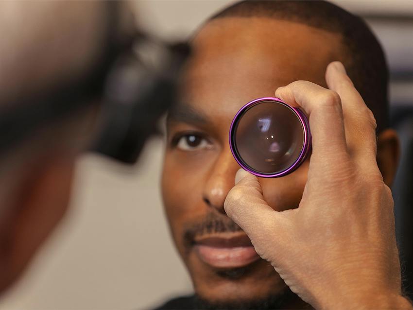
Injuries, infections, chemical exposures, and other forms of corneal trauma can leave behind scarring that permanently affects vision. We evaluate and manage corneal scarring across the full spectrum of severity, from observation and optical management through to surgical intervention, tailoring the approach to the nature and extent of each guest's condition.
Post-Surgical Surface Services
Post-Surgical Surface Services
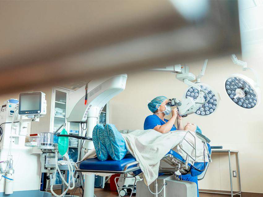
Dry eye, epithelial irregularity, persistent defects, and other surface complications following eye surgery are more common than most guests realize, and they are very treatable when addressed by the right team. We work with guests whose recovery hasn't gone as expected to help them achieve the outcomes their original procedures were meant to deliver.

Dry Eye & Ocular Surface Disease
Dry Eye & Ocular Surface Disease
Our approach to dry eye and ocular surface disease is comprehensive, covering the full spectrum from mild situational dry eye to severe, refractory disease driven by systemic conditions, prior surgery, or complex combinations of underlying factors. Every guest receives a thorough evaluation and a treatment plan built around what the findings actually show.





Why Choose Boling Vision Center?
Boling Vision Center's Cornea & Ocular Surface Center offers the diagnostic depth, therapeutic breadth, and clinical expertise that complex corneal and ocular surface conditions demand. With four locations across Northern Indiana, an on-site Joint Commission-certified surgery center, and a team known throughout the region for taking difficult cases seriously, we are the practice guests and referring physicians turn to when they need answers, options, and results. Over 65 years of community trust, and counting. Amazing things happen here.
Cornea & Ocular Surface Center Frequently Asked Questions
What kinds of conditions does the Cornea & Ocular Surface Center treat?
We treat the full spectrum of corneal and ocular surface conditions, including dry eye disease, meibomian gland dysfunction, blepharitis, keratoconus and other ectatic conditions, corneal scarring and trauma, inflammatory and autoimmune-related eye disease, post-surgical surface complications, and corneal dystrophies and degenerations. If a condition affects the front surface of the eye, it falls within our scope of care.
How is the Cornea & Ocular Surface Center different from a general eye exam?
A comprehensive eye exam evaluates overall eye health and determines refractive needs. The Cornea & Ocular Surface Center goes significantly deeper, using specialized diagnostic tools to evaluate the tear film, meibomian glands, corneal structure, and ocular surface health in detail and build targeted treatment plans based on those specific findings. The clinical focus, diagnostic toolkit, and range of treatment options available are considerably more extensive than what a routine exam provides.
Do I need a referral to be seen at the Cornea & Ocular Surface Center?
No referral is required. You're welcome to schedule directly at any of our four Northern Indiana locations. If you've been referred by another provider, we're happy to coordinate with them and keep them informed as part of your care team.
Can the Cornea & Ocular Surface Center help if I've seen other providers without improvement?
We hope so, and it's one of the most common reasons patients come to us. Boling Vision Center is known throughout the region as a practice that other providers refer to when they need answers on complex cases. A fresh evaluation with our full diagnostic toolkit may reveal factors that weren't identified or addressed in prior care, and our range of treatment options means we can offer approaches that may not have been available elsewhere.
What should I expect at my first Cornea & Ocular Surface Center appointment?
Your first appointment will be more thorough and time-intensive than a routine eye exam. Depending on your condition, it may include corneal imaging, tear film analysis, meibomian gland assessment, and a detailed slit-lamp evaluation, in addition to a full review of your ocular and medical history. Plan for an appointment of roughly one to one and a half hours. Bring a list of current medications, any prior eye care records or imaging, and your current glasses and contact lenses. If you wear contacts, removing them for several days before your appointment can improve the accuracy of corneal imaging.