Protecting the Vision That Matters Most

Central vision, the sharp, detailed sight we use for reading, recognizing faces, driving, and engaging with the world around us, depends entirely on the health of a small but critical area of the retina called the macula. When AMD affects the macula, that central vision becomes distorted, blurred, or lost, often gradually and silently before the changes are fully noticed. The encouraging reality is that AMD, particularly in its earlier stages, is very manageable with the right monitoring and treatment. The earlier it is detected, the more options are available and the better the long-term outcome. Schedule a macular evaluation at any of our four Northern Indiana locations today.

What Is Age-Related Macular Degeneration?

The macula is a small, specialized area at the center of the retina responsible for the sharp, detailed central vision we rely on for virtually every visually demanding task. In AMD, the macula gradually deteriorates, disrupting central vision while typically leaving peripheral vision intact. AMD is primarily a disease of aging. The risk increases significantly after age 60 and continues to rise with each decade.

While AMD rarely causes complete blindness because peripheral vision is preserved, it can cause substantial visual disability that significantly affects independence and quality of life. AMD is one of the most important conditions we screen for during comprehensive eye exams, because early detection allows for intervention at a stage when treatment is most effective.

Banner media

Types of AMD

AMD presents in two distinct forms, each with different characteristics, progression patterns, and treatment approaches.

Dry AMD

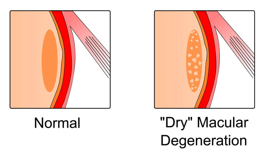
Dry AMD is the most common form, accounting for the majority of AMD cases. It develops as the macula gradually thins with age, and small deposits called drusen accumulate beneath the retinal tissue. These changes occur slowly, often over years, and may produce no noticeable symptoms in the early stages.

As dry AMD progresses, guests may notice a gradual blurring or dimming of central vision, difficulty reading in low light, and a fading of color vibrancy. In advanced stages, dry AMD can progress to geographic atrophy, in which large areas of retinal cells are lost, causing significant central vision impairment.

Dry AMD can also progress to wet AMD in some guests, making regular monitoring essential regardless of which stage is present.

Dry Macular Degeneration graphic

Wet AMD

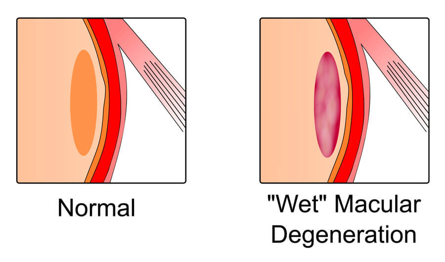
Wet AMD is less common but more rapidly progressive and more immediately threatening to central vision. It occurs when abnormal blood vessels grow beneath the retina in a process called choroidal neovascularization. These vessels are fragile and prone to leaking blood and fluid, which damages the light-sensitive cells of the macula.

Wet AMD typically produces more sudden and dramatic vision changes than dry AMD, including distortion that makes straight lines appear wavy, dark or blank spots in the central visual field, and rapid deterioration of reading and detail vision. Without prompt treatment, the abnormal vessels scar, causing permanent central vision loss.

Any guest with dry AMD who notices a sudden change in vision or new distortion should seek evaluation immediately, as this may signal conversion to wet AMD.

Wet AMD graphic

Megan Heil

Risk Factors for AMD

While age is the primary risk factor for AMD, several additional factors increase the likelihood of developing the condition or accelerating its progression:

  • Age: Risk increases significantly after 60 and continues to rise with each decade.
  • Family history: A first-degree relative with AMD meaningfully raises the risk.
  • Smoking: One of the most significant modifiable risk factors; smokers have a substantially higher risk of developing AMD and of faster progression.
  • Cardiovascular disease: High blood pressure, elevated cholesterol, and other cardiovascular risk factors are associated with increased AMD risk.
  • UV exposure: Cumulative ultraviolet light exposure over a lifetime contributes to retinal oxidative stress.
  • Diet and nutrition: Diets low in antioxidants and omega-3 fatty acids are associated with higher AMD risk; the AREDS2 nutritional supplement formula has been shown to reduce the risk of progression in intermediate AMD.
  • Race: AMD is more common in Caucasian populations than in other racial groups.
    Understanding your personal risk profile is one of the reasons regular comprehensive eye exams are so important, even in the absence of symptoms.

Awesome staff! My daughter is 2; they treated her and me with respect and worked really well with her.

Karma Helvey
Quote Background

Symptoms of AMD

AMD often develops silently before producing obvious symptoms, particularly in its early stages. Signs that may indicate AMD include:

  • Blurry, hazy, or dim central vision
  • Difficulty reading, particularly in lower-light conditions
  • Straight lines appearing wavy or distorted
  • A dark, blurry, or blank spot in the center of the visual field
  • Colors appearing less vivid or washed out
  • Increased difficulty recognizing faces
  • Needing brighter light for reading and detailed tasks

Many guests with early AMD are unaware of any vision change, which is why regular dilated retinal examinations and advanced imaging are the most reliable means of early detection.

Banner media

How We Diagnose AMD

Early and accurate diagnosis of AMD requires more than a standard eye exam. At Boling Vision Center's Medical Retina Center, we use a comprehensive suite of advanced diagnostic tools to detect AMD at its earliest stages and characterize its extent and activity with precision.

Dilated Retinal Examination

A thorough dilated examination of the retina allows our providers to directly visualize the macula, assess the presence and extent of drusen, identify areas of retinal pigment epithelium changes, and evaluate for signs of choroidal neovascularization.

Optical Coherence Tomography (OCT)

OCT provides detailed cross-sectional imaging of the retinal layers, allowing us to detect subtle structural changes in the macula that may not be apparent on clinical examination alone. It is an essential tool for both diagnosis and ongoing monitoring, providing a level of structural detail that significantly enhances our ability to track disease activity and treatment response.

OCT Angiography (OCTA)

OCTA is a non-invasive imaging modality that visualizes the retinal and choroidal vasculature without the need for dye injection. It allows us to detect early choroidal neovascularization and monitor its extent and activity, providing critical information for treatment planning in wet AMD.

Fluorescein Angiography

In select cases, fluorescein angiography, which involves the injection of a fluorescent dye to image blood flow in the retina and choroid, provides additional detail about the location and activity of abnormal vessels in wet AMD.

Home Monitoring

For guests with intermediate AMD or early wet AMD who are between office visits, home monitoring tools, including the Amsler grid and digital monitoring devices, allow for self-assessment of central vision and early detection of changes that warrant prompt evaluation.

AMD Treatment Options

Treatment for AMD depends on the type and stage of the condition. At Boling Vision Center, we offer the full range of current AMD treatment options and stay current with advances in this rapidly evolving field.

Monitoring and Lifestyle Management for Early Dry AMD

For guests with early dry AMD, the primary approach is regular monitoring combined with lifestyle modifications and nutritional supplementation designed to slow progression. These include smoking cessation, UV protection, cardiovascular risk factor management, and AREDS2 nutritional supplementation for guests with intermediate AMD, which has been shown in large clinical trials to reduce the risk of progression to advanced AMD.

Anti-VEGF Injections for Wet AMD

The primary treatment for wet AMD is intravitreal injection of anti-VEGF (vascular endothelial growth factor) agents, which block the growth and leakage of the abnormal blood vessels responsible for the vision-threatening damage of wet AMD. These injections are administered in the office and are generally well tolerated.

Anti-VEGF therapy has transformed the treatment of wet AMD, converting what was once a condition of near-certain severe vision loss into one that can be stabilized and, in many cases, improved with appropriate treatment. Early initiation of treatment produces the best outcomes, reinforcing the importance of prompt evaluation when any sudden vision change occurs.

Valeda Light Therapy for Dry AMD

Boling Vision Center is among a select group of practices offering Valeda Light Delivery System, a photobiomodulation treatment for dry AMD. Valeda uses specific wavelengths of light delivered to the retina to support the health and function of retinal cells and has shown promising results in slowing the progression of dry AMD and supporting visual function.

Valeda is a non-invasive, in-office treatment that represents a meaningful addition to the dry AMD management toolkit for eligible guests. Ask our team whether Valeda may be appropriate for your situation.

Background media

Living With AMD

An AMD diagnosis can feel daunting, but it is important to understand that most guests with AMD, particularly those diagnosed early and managed appropriately, maintain meaningful central vision for many years.

Proactive management, consistent follow-up, and honest communication with your care team are the most powerful tools available for preserving vision and quality of life with AMD. 

Our team is committed to staying closely engaged with every AMD guest, monitoring for progression, adjusting treatment as the condition evolves, and ensuring that every guest has the information and support they need to navigate the condition with confidence.

Low vision resources and referrals are also available for guests whose AMD has progressed to a point where additional support is beneficial.

Why Choose Boling Vision Center?

Boling Vision Center's Medical Retina Center combines advanced diagnostic imaging, the full range of AMD treatment options, including Valeda light therapy, and providers with the clinical depth to manage AMD at every stage with precision and genuine care. With four Northern Indiana locations, over 65 years of trusted community care, and a commitment to staying at the forefront of retinal medicine, we're the practice Michiana trusts to protect the vision that matters most. Amazing happens here.

Richard Boling II

Frequently Asked Questions Age-Related Macular Degeneration

Can AMD be cured?

There is currently no cure for AMD, but it is very manageable, particularly when detected early. Dry AMD can be slowed significantly with appropriate monitoring, lifestyle modifications, and nutritional supplementation. Wet AMD can be stabilized and often improved with anti-VEGF injections. The goal of treatment is to preserve as much central vision as possible for as long as possible.

Will AMD make me go blind?

AMD affects central vision but typically spares peripheral vision, meaning complete blindness from AMD alone is uncommon. However, advanced AMD can cause significant central vision loss that substantially affects the ability to read, drive, and recognize faces. Early detection and consistent management are the most effective means of preventing that outcome.

How often do I need to be seen if I have AMD?

The frequency of monitoring depends on the stage and type of AMD. Early dry AMD may require monitoring every six to twelve months. Intermediate AMD typically warrants more frequent evaluation. Guests receiving anti-VEGF treatment for wet AMD are seen at regular intervals determined by their treatment protocol and disease activity. Your provider will recommend the appropriate schedule for your individual situation.

Are there nutritional supplements that help with AMD?

Yes. The AREDS2 formula, which contains specific doses of vitamins C and E, lutein, zeaxanthin, zinc, and copper, has been shown in large clinical trials to reduce the risk of progression from intermediate to advanced AMD. It is not a treatment for early AMD and is not proven to prevent AMD from developing, but it is an important tool for appropriate candidates. Ask your provider whether AREDS2 supplementation is recommended for your situation.

What should I do if I notice sudden changes in my central vision?

Seek evaluation immediately. Sudden changes in central vision, new distortion, or a new dark spot in the visual field can signal conversion from dry to wet AMD or another serious retinal condition requiring prompt treatment. Do not wait for a scheduled appointment if you notice these changes.

Is AMD hereditary?

AMD has a significant hereditary component. Having a first-degree relative with AMD meaningfully increases your risk, and certain genetic variants are associated with higher AMD susceptibility. Guests with a family history of AMD should begin regular retinal screening earlier and more frequently than the general population.

Amazing Happens Here

Three generations. One unwavering standard.

Contact Us
Accessibility: If you are vision-impaired or have some other impairment covered by the Americans with Disabilities Act or a similar law, and you wish to discuss potential accommodations related to using this website, please contact our Accessibility Manager at (574) 293-3545.
Contact Us