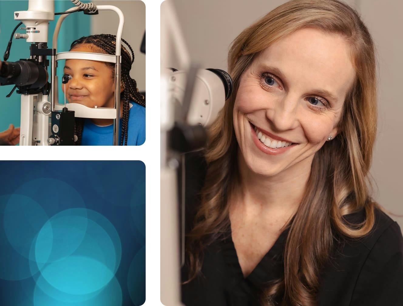
The cornea and ocular surface are complex, and the conditions that affect them often raise more questions than a single appointment can answer.
Corena & Ocular Surface FAQ's


Answers to Your Cornea & Ocular Surface Questions
Whether you're navigating a new diagnosis, weighing treatment options, or simply trying to understand what's happening with your eyes, having clear and honest information makes all the difference. This page brings together answers to the questions we hear most often across the full range of cornea and ocular surface conditions we treat at Boling Vision Center. If your question isn't covered here, our team is always glad to help. Schedule a consultation at any of our four Northern Indiana locations and let's talk through what's on your mind.
Frequently Asked Questions General Cornea & Ocular Surface Health
What does the cornea actually do?
The cornea is the clear, dome-shaped tissue at the very front of the eye. It serves two essential functions: it acts as a protective barrier against the outside world, and it provides roughly two-thirds of the eye's total focusing power. Because light must pass through the cornea before reaching the lens and retina, even subtle changes in its clarity, curvature, or surface quality can have a significant impact on vision.
What is the ocular surface, and why does it matter?
The ocular surface refers to the entire front surface ecosystem of the eye, including the cornea, conjunctiva, eyelids, and tear film. These structures work together as an integrated system to keep the eye clear, comfortable, lubricated, and protected. When any part of that system is disrupted, whether by disease, injury, inflammation, or environmental factors, the effects are felt across the whole system. Good ocular surface health is foundational to good vision.
How do I know if I have a corneal problem?
Common signs that something may be affecting the cornea or ocular surface include blurry or distorted vision that doesn't fully correct with glasses, increased sensitivity to light or glare, a persistent foreign body sensation, redness or discomfort that doesn't resolve, visible cloudiness or haziness on the front of the eye, and frequent prescription changes. That said, some corneal conditions, including early keratoconus and certain dystrophies, can be present without obvious symptoms, which is one reason regular comprehensive eye exams matter.
Can corneal conditions affect both eyes?
Yes. Many corneal conditions, including keratoconus, Fuchs' endothelial dystrophy, and dry eye disease, commonly affect both eyes, though they may progress at different rates in each. Some conditions, particularly those resulting from injury or localized infection, may initially affect only one eye. Your provider will evaluate both eyes thoroughly, regardless of where symptoms are most apparent.
Frequently Asked Questions Dry Eye & Ocular Surface Disease
What is the difference between dry eye and ocular surface disease?
Dry eye disease is one type of ocular surface disease, but the two terms aren't interchangeable. Ocular surface disease is a broader category that includes any condition affecting the health and function of the cornea, conjunctiva, eyelids, or tear film. Dry eye is the most common of these conditions, but ocular surface disease also encompasses conditions like blepharitis, meibomian gland dysfunction, ocular rosacea, and more serious inflammatory and autoimmune conditions that affect the surface.
Why haven't over-the-counter drops helped my dry eye?
Over-the-counter artificial tears address the symptom of dryness by temporarily supplementing the tear film, but they don't address the underlying cause of why the tear film is unstable in the first place. For many guests, dry eye is driven by meibomian gland dysfunction, inflammation, or other factors that drops alone can't resolve. A thorough dry eye evaluation identifies what's actually driving the condition, which is the starting point for treatment that produces lasting improvement.
Is dry eye related to screen use?
Screen use is a contributing factor for many people. When focusing on a screen, blink rate decreases significantly, which reduces the regular distribution of tears across the ocular surface and accelerates evaporation. While screen use alone is unlikely to cause dry eye disease in someone with a completely healthy ocular surface, it commonly worsens symptoms in those who are predisposed. Managing blink habits and screen ergonomics is a useful part of an overall dry eye management plan, though rarely sufficient on its own for moderate to severe cases.
Can dry eye affect my vision?
Yes. An unstable tear film disrupts the smooth optical surface of the cornea, which can produce blurry or fluctuating vision, reduced contrast sensitivity, and difficulty with tasks like reading, driving, or using screens. For some guests, the visual impact of dry eye is as significant as the discomfort, and treating the underlying condition often produces meaningful improvements in vision quality alongside symptom relief.
Frequently Asked Questions Keratoconus & Corneal Shape Conditions
What is the difference between keratoconus and astigmatism?
Astigmatism is an extremely common refractive condition caused by an irregularly shaped cornea or lens, and it's easily corrected with glasses or standard contact lenses. Keratoconus is a progressive disease in which the cornea gradually weakens and bulges into a cone shape, producing a form of irregular astigmatism that becomes increasingly difficult to correct with conventional lenses as it advances. Not all astigmatism is keratoconus, but keratoconus does produce astigmatism; the distinction matters because the management is very different.
If I have keratoconus, will I eventually need a corneal transplant?
The majority of guests with keratoconus never progress to needing a corneal transplant. With early diagnosis, appropriate monitoring, and timely intervention, including corneal cross-linking when indicated, most guests maintain good functional vision throughout their lives with specialty contact lenses. Transplantation becomes a consideration only in cases of advanced disease where functional vision can no longer be achieved through other means.
What is corneal ectasia, and how is it different from keratoconus?
Corneal ectasia is a broader term for any condition involving progressive corneal thinning and bulging, of which keratoconus is the most common form. Post-surgical ectasia, which can occur as a rare complication of corneal refractive surgery, is another form. The underlying mechanism is similar: weakening of the corneal collagen structure allows the cornea to deform under normal intraocular pressure. Management approaches, including cross-linking, also overlap significantly between these conditions.
Frequently Asked Questions Corneal Infections & Injuries
What should I do if I get something in my eye?
For minor surface debris, gentle flushing with clean water or sterile saline is appropriate. Do not rub the eye, as this can drive a foreign body deeper or cause additional surface damage. If the sensation persists after flushing, if vision is affected, or if there is significant pain or redness, seek care promptly. For chemical exposures, flush immediately and continuously with water for at least 15 to 20 minutes before seeking emergency evaluation; don't wait to see if symptoms improve.
How serious is a corneal infection?
Corneal infections, also called infectious keratitis, can be very serious and should always be evaluated promptly. Bacterial keratitis in particular can progress rapidly, and delays in treatment increase the risk of corneal scarring and permanent vision loss. Contact lens wearers are at higher risk and should seek same-day evaluation for any red, painful eye, particularly if accompanied by discharge or sensitivity to light.
Can a scratched cornea heal on its own?
Most superficial corneal abrasions heal within 24 to 72 hours without leaving lasting damage, as the corneal epithelium regenerates quickly. Larger or deeper abrasions may take longer and benefit from a protective bandage contact lens and lubricating drops to support healing and reduce discomfort. Any abrasion that doesn't improve within a couple of days, or that is accompanied by significant pain, discharge, or worsening vision, warrants evaluation to rule out infection or deeper injury.
Frequently Asked Questions Corneal Surgery & Procedures
What is the recovery like after corneal surgery?
Recovery varies considerably depending on the procedure. Surface treatments like phototherapeutic keratectomy typically involve a healing period of one to two weeks for the epithelium to regenerate, with some light sensitivity and blurry vision during that time. Endothelial transplants like DMEK often produce good functional vision within weeks to a few months. Full-thickness corneal transplants involve the longest recovery, with visual rehabilitation that can extend over a year as the cornea heals and the prescription stabilizes.
How long does a corneal transplant last?
Modern corneal transplants, particularly endothelial transplants like DMEK, have excellent long-term survival rates. Full-thickness grafts can last decades in many cases. Graft survival depends on a number of factors, including the underlying condition, the health of the recipient eye, adherence to post-operative care, and the prompt treatment of any rejection episodes. Regular long-term follow-up is important for all transplant recipients.
Can I drive after corneal surgery?
Not on the day of the procedure. Specific driving restrictions vary depending on the type of surgery and how quickly vision recovers. Your provider will give you clear guidance on when it's safe to resume driving based on your individual recovery progress.
Frequently Asked Questions Finding the Right Care
Do I need a referral to be seen at Boling Vision Center's Cornea & Ocular Surface Center?
No referral is required. You're welcome to schedule directly at any of our four Northern Indiana locations. That said, if you've been referred by another provider, we're happy to coordinate with them and keep them informed as part of your care team.
What should I bring to my first cornea or ocular surface appointment?
Bring a list of all current medications, including eye drops; any prior eye care records or imaging if available; your current glasses and contact lenses; and your insurance information. If you wear contact lenses, it's helpful to remove them for several days before your appointment if possible, as contact lens wear can temporarily alter corneal shape and affect imaging accuracy.
I've seen other providers and haven't gotten answers. Can Boling Vision Center help?
We hope so. Boling Vision Center is known throughout the region as the practice that other providers refer to when they need answers on complex cases. Our team takes the time to look at the full picture, ask the right questions, and pursue a diagnosis rather than simply managing symptoms. If you've been told your condition is unclear or that nothing more can be done, we'd encourage you to come in for a second opinion.
Why Choose Boling Vision Center?
Boling Vision Center's Cornea & Ocular Surface Center offers the diagnostic depth, treatment breadth, and genuine attentiveness that complex corneal and ocular surface conditions demand. With four locations across Northern Indiana, an on-site accredited surgery center, and a team known for taking difficult cases seriously, we're the practice guests and referring physicians turn to when they need answers and results. Amazing happens here.